

Skin & Regeneration
Changes in Red Lesions and Pore Visibility Were Reported After RF Microneedling with NAD+
A November 8, 2026 presentation reported changes in red lesions, skin texture, pore visibility, and skin tone after combining an NAD+ (nicotinamide adenine dinucleotide) formulation with radiofrequency (RF) microneedling. Three cases with clearer changes were selected from six people who received two combined sessions in the speaker’s own practice. The speaker disclosed a Vitamedical-related conflict of interest on the slides. These small, uncontrolled observations had no quantitative scores and cannot isolate NAD+’s contribution. The next question is whether the formulation differs from a control matched for other ingredients and properties when both accompany the same RF procedure.
ASLS 2026 talk series: ① Evidence and limits · ② Talk findings and limits of uncontrolled observations (this article) · ③ At the ASLS 2026 talk.
About this article
This article explains a conference presentation and related literature; it does not recommend a particular procedure. The site editorial team did not author the cited studies. This article does not assess authorization, classification, or indications in Korea.
Presentation information
- Event: ASLS KOREA 2026 AUTUMN CONGRESS, November 7–8, 2026, COEX, Samseong-dong, Seoul.
- Presentation: November 8, 2026, scheduled 15:10–15:25 in D Room; a 15-minute lecture opening the session Pore Treatment & Beyond, Plus Hair Removal.
- Official registered title: Remodeling Pores and Post-Acne Skin with an NAD+ Bioactive Platform.
- Speaker: Hyojung Moon, MD, director of Beyond Clinic. The presentation used 19 English-language slides.[1]
- The presentation materials label the NAD+ formulation as Revergen and the accompanying device as Potenza.
Key points
- The November 8, 2026 presentation showed three selected cases from six receiving two combined NAD+ formulation and RF microneedling sessions. Changes in red lesions, texture, pore visibility, and tone were reported.
- The three cases with clearer changes were selected. The presentation also reported that skin tone appeared brighter in the other three cases.
- Assessment relied on photographs and physician observation, without quantitative scores. It cannot establish an overall response rate or magnitude of change.
- Without a comparator, the contributions of RF and the formulation cannot be separated. A trial using the same RF with the NAD+ formulation or a matched vehicle was proposed.
Where the presentation took place
Hyojung Moon, MD, presented observations from her own practice at ASLS KOREA 2026 AUTUMN CONGRESS, held at COEX in Samseong-dong, Seoul, on November 8, 2026. The 15-minute lecture was scheduled for 15:10–15:25 as the opening talk in the D Room session Pore Treatment & Beyond, Plus Hair Removal.
The official title was Remodeling Pores and Post-Acne Skin with an NAD+ Bioactive Platform. Remodeling refers to tissue reorganization. The title does not mean that biopsy measurements established this process in the presented cases.[1]
The starting question: metabolic support during skin repair
The presentation asked whether the metabolic processes needed for repair could also be supported after a procedure stimulates skin. NAD+ is a coenzyme, a substance used alongside enzymes in cellular energy metabolism and responses to damage.[2]
NAD+ takes part in oxidation–reduction, or redox reactions, that transfer electrons. It is also involved in responses to damage in DNA, the genetic material, and in stress adaptation. These biological roles do not establish what an external formulation does in skin.[1,2]
Niacinamide studies as background
For background, the presentation reviewed skin studies of niacinamide, a form of vitamin B3. This precursor goes through intermediate steps in NAD+ synthesis; it is a different molecule from NAD+.[2]
A niacinamide pore study reported image-based changes from baseline, which must be distinguished from superiority over vehicle, the comparison formulation without niacinamide.[3] An acne trial’s failure to detect a difference between niacinamide and an antibiotic gel also does not establish equivalence.[4] The acne source recorded minor adverse events in 14 of 40 nicotinamide recipients and 11 of 40 clindamycin recipients.[4] A pigmentation study reported less hyperpigmentation on the niacinamide side than on the vehicle side; reduced pigment transfer was observed in separate cell experiments.[5] None of these findings establishes a result for the current NAD+ formulation.
NAD+ deficiency in pore- or acne-affected skin remains a hypothesis
The presentation proposed a hypothesis for skin with overlapping sebum, redness, pigment marks, barrier damage, and scars: inflammation and damage responses might change NAD+ demand and consumption.[1]
Studies have linked NAD+ to repair and damage responses in aged mouse skin, skin explants, and ultraviolet-exposed skin cells.[6,7] The literature reviewed for the presentation did not provide direct measurements of NAD+ deficiency in human pore- or acne-affected skin. This literature does not justify labeling an individual patient’s skin NAD+-deficient.
The combination hypothesis depends on delivery
By combining small needles with RF energy, RF microneedling produces limited injury intended to stimulate tissue change. The combination hypothesis asks whether metabolic support could accompany that stimulus.[1]
The hypothesis depends on the formulation reaching viable skin cells in a usable form. Experiments with other substances have reported increased delivery to the outer skin layer, as well as failure to observe dermal delivery under particular conditions.[8,9] Actual delivered amounts and intracellular NAD+ changes were not measured in these cases.[1]
How the six cases were observed
The six people described in the presentation had two combined full-face sessions of RF microneedling and the NAD+ formulation, 2 weeks apart, with follow-up at 2-week intervals. The formulation volume was listed as 4 mL per session. The amount of NAD+ reaching cells was not measured.[1]
Assessment used three-dimensional facial imaging and physician observation, but the presentation reported no quantitative scores for lesion counts, pore area or volume, or pigmentation indices.[1]
Of the six cases, three with clearer changes were selected for presentation. They cannot represent a group average or an individual’s expected outcome. The time between the final session and final assessment was not consistently expressed in relative weeks.[1]
The three cases shown
Case 1: persistent red, papule-like lesions on both cheeks after an earlier injectable procedure
The first case involved persistent red, papule-like lesions on both cheeks after an earlier injectable procedure, as described in the presentation. Papule-like refers to a small raised appearance; it does not confirm a diagnosis. After two sessions, distinct changes in red marks and texture relative to baseline were reported.
The presentation acknowledged that the change was less distinct in photographs than in person.[1]
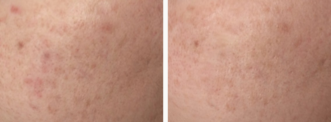
Case 1, cropped cheek skin: left, baseline; right, after two sessions. Eyes, nose, mouth, ears, facial outline, and capture dates were concealed. The photographs were taken approximately 24 days apart. Lighting and skin-surface gloss differed between captures.
Case 2: long-standing inflammatory acne
In the second case, the presentation described red, raised lesions and pustules on the cheeks, around the mouth, and on the chin, together with surrounding redness. Pustules are lesions containing pus.
After two sessions, the presentation reported fewer new breakouts and a distinct change in texture relative to baseline. It also reported fewer active inflammatory lesions and less surrounding redness, with some post-inflammatory marks remaining.[1]
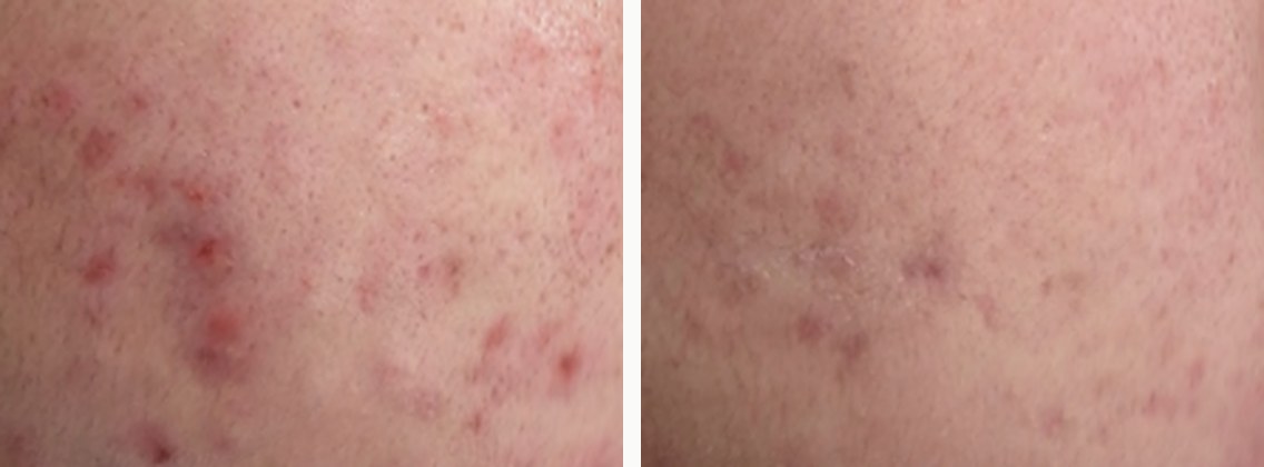
Case 2, cropped cheek skin: left, baseline; right, after two sessions. Eyes, nose, mouth, ears, facial outline, and capture dates were concealed. The photographs were taken approximately 64 days apart. Lighting and skin-surface gloss differed between captures.
Case 3: visible pores and uneven skin tone
For the third case, the presentation described visible pores on the cheeks and nose, uneven tone, and scattered pigment marks. It reported that, after two sessions, cheek pores appeared less prominent than at baseline, skin tone appeared brighter, and pigment marks appeared lighter.[1]
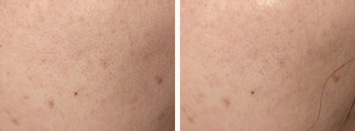
Case 3, cropped cheek skin: left, baseline; right, after two sessions. Eyes, nose, mouth, ears, facial outline, and capture dates were concealed. The photographs were taken approximately 19 days apart. Lighting and skin-surface gloss differed between captures.
Three patterns described in the presentation
The presentation organized the cases into the patterns below; these groupings do not measure the mechanism behind any change.[1]
| Pattern described | Change reported in the presentation | Interpretation limit |
|---|---|---|
| Persistent red lesions and texture after an earlier procedure | Less visible red papule-like marks and a change in texture | No confirmed lesion diagnosis or isolated ingredient contribution |
| Inflammatory acne | Fewer new lesions and a change in texture | No counted lesion outcome quantifying the change |
| Visible pores and uneven tone | Pores appeared less visible and skin tone appeared brighter | No quantitative measurement of anatomical pore size or pigment amount |
According to the presentation, skin tone also appeared brighter in the other three cases. Without individual courses or magnitudes of change, this cannot be presented as a quantitative result for all six.[1]
The closest published evidence discussed
The presentation placed these observations alongside human NAD+ skin studies and trials that added a substance to the same RF procedure. It reported finding no controlled trial that directly matched this combination in its literature review.[1]
Studies applying NAD+ to human skin
In a single-arm, uncontrolled study of 36 women with melasma, another NAD+ formulation was applied after ordinary microneedling. At the reported week-21 assessment, the mean Melasma Area and Severity Index (MASI) fell from 16.8 at baseline to 6.9.[10] No RF was used, and the contributions of microneedling, photoprotection, time, and NAD+ could not be separated. A melasma score cannot be assigned to these pore and acne observations.
An exploratory psoriasis study involved 37 patients in total. In a separate comparison involving 11, NAD+ ointment and vehicle were applied to similar plaques within the same person. Reported plaque scores at 4 weeks were 5.8 and 7.2, respectively; lower scores indicated less severe plaques (p = 0.0294).[11] The presentation noted no significant difference in comparisons with anthralin ointment, a topical psoriasis treatment, and that randomization was not stated. Psoriasis, the formulation, and the application method differ from these pore and acne observations.[1,11]
Trials adding another substance to the same RF procedure
A randomized split-face trial in 24 people with atrophic acne scars, depressed scars after acne, used the same RF on both sides, with poly-L-lactic acid (PLLA) on one side and sterile water on the other. At 6 months, the reported between-side difference in texture change was −1.18, favoring the PLLA side (p = 0.045).[12] There were multiple primary outcomes, the p value was borderline, and actual delivery was not measured. Another ingredient’s combination result cannot be assigned to NAD+. The authors described adverse events as infrequent, transient, and self-limited, with no serious complications reported in that trial.[12]
In contrast, a randomized split-face melasma trial analyzed 26 of 30 participants. Both sides received the same RF, with a combination formulation containing 0.3% polynucleotide (PN) on one side and saline on the other. No added benefit was detected in the between-side modified Melasma Area and Severity Index (mMASI) comparison through 6 months (p = 0.784).[13] Adding a substance does not necessarily produce an additional clinical benefit.
These studies can help shape the next comparison, while leaving the independent effect of the presented combination unresolved.
How to interpret the reported changes
After the combination, the presentation reported changes in red lesions, texture, pore visibility, and tone. Explanations based on repair, inflammation, or tissue reorganization remain interpretive hypotheses because the relevant molecular pathways and tissue changes were not measured directly.[1]
The presentation identified these limitations:
- Six uncontrolled cases: without a comparison group or a contralateral control, their course without the combination is unknown.
- Three selected cases shown: cases with clearer changes cannot represent the distribution of outcomes across everyone treated.
- Photograph-based assessment without quantitative scores: the magnitude and reproducibility of change remain unknown.
- Short follow-up and varying baseline dates: persistence and delayed reactions cannot be determined.
- Different lighting and skin-surface gloss between captures: these can influence the appearance of pores, texture, and tone.
- Intracellular delivery was not measured: linking appearance to increased intracellular NAD+ or a particular enzyme’s activity remains a hypothesis.
- NAD+’s contribution cannot be isolated: there was no RF-only comparison to separate the formulation’s contribution.[1]
The contributions of the diluent, additives, and time were not separated, and accompanying treatments were not described. An acne randomized trial comparing RF microneedling with another device also reported lesion changes without NAD+.[14] Its device and conditions differ, so its effect size cannot be transferred here. It nonetheless shows why the observed changes cannot all be attributed to NAD+.
Adverse-event data were not presented for these cases. Unreported data cannot be converted into a finding that no events occurred. Records need to cover all participants’ follow-up, reaction onset and duration, severity, and any treatment.
The separate uncontrolled melasma study reported pain, along with redness and swelling that lasted 24–48 hours. Post-inflammatory hyperpigmentation (PIH), dark marks that can follow inflammation, was not reported in that small study.[10] Neither that finding nor another ingredient’s trial establishes the safety of this combination. Repeated local exposure, rare delayed reactions, and drug interactions remain insufficiently characterized in the literature synthesis.
An FDA safety notice describes serious RF microneedling injuries involving burns, scars, fat loss, disfigurement, and nerve damage. These are not events reported for these six patients or an incidence estimate. The procedure is not for home use.[15]
The FDA has also reported severe chills, shaking, vomiting, and fatigue after compounded injectable NAD+, sometimes requiring medical treatment, and warned about unsuitable food-grade ingredients, microbes, and endotoxins. Those preparation-quality concerns do not establish contamination of this formulation. Ingredient and diluent composition, route suitability, reconstitution stability, sterility, and endotoxin control require separate verification.[16]
What the next study should compare
The proposed comparison asks whether the same RF produces an additional change when accompanied by the NAD+ formulation rather than a formulation lacking NAD+. The proposal brings together the presentation’s suggestion and design principles from the literature. It is not a fixed treatment schedule or an established trial plan.
Starting with participants who have similar active acne would make the comparison easier to interpret. Persistent red lesions after an earlier procedure, active acne, and concerns limited to pores and tone should not be combined into one primary outcome.
In a split-face design, both sides would receive the same RF. The side receiving the NAD+ formulation would be randomly assigned; the other would receive a matched vehicle, with NAD+ omitted and other ingredients and properties matched. Actual composition needs confirmation before matching diluent, additives, acidity, osmotic pressure, viscosity, and volume. If a matched vehicle cannot be obtained and the comparator is RF alone, the study would estimate the whole formulation’s added contribution, not NAD+ alone.
Assessors would be blinded to the assigned sides. The primary outcome would be set in advance; for example, the within-person difference in inflammatory lesion reduction 12 weeks after the final session. Pores, pigmentation, and redness would be assessed separately, rather than combined into a new score. Lighting, posture, cleansing conditions, and accompanying treatments also need prespecification.
Results should give each person’s paired difference and its uncertainty, with the two facial sides never counted as independent people. Sample size and session intervals require decisions based on the endpoint, variability, and safety review.
Separate mechanistic studies would need to measure the actual formulation’s stability, exposure in each skin layer, and intracellular versus extracellular material and breakdown products. Before implementation, composition, route, reconstitution quality, comparator, applicable review and consent, and trial registration need confirmation.
Conflict-of-interest disclosure
The speaker disclosed Vitamedical (C) on the slides.[1] The meaning of C has not been confirmed. The cases are uncontrolled observations from the speaker’s own practice, and the speaker is also this article’s medical reviewer.
What this presentation did not establish
The presentation did not assess the effect of NAD+ alone, deficiency and replenishment in human pore- or acne-affected skin, new collagen formation, or scar-tissue reorganization.[1] It also did not establish comparative superiority, long-term safety, or an individual’s expected result.
This article explains the reported changes and limits of the combination observations. Formulation and device names identify labels in the presentation materials; they are not recommendations for a product or procedure. Seek medical assessment for persistent redness or accompanying pain.
References
- Moon H. Remodeling Pores and Post-Acne Skin with an NAD+ Bioactive Platform. ASLS KOREA 2026 AUTUMN CONGRESS; November 8, 2026; COEX, Seoul, D Room. Conference presentation in English, 19 slides. Public release of the complete deck has not been decided.
- Covarrubias AJ, Perrone R, Grozio A, Verdin E. NAD+ metabolism and its roles in cellular processes during ageing. Nat Rev Mol Cell Biol. 2021;22(2):119-141. PubMed.
- Chiu PC, Chan CC, Lin HM, Chiu HC. The clinical anti-aging effects of topical kinetin and niacinamide in Asians: a randomized, double-blind, placebo-controlled, split-face comparative trial. J Cosmet Dermatol. 2007;6(4):243-9. PubMed.
- Khodaeiani E, Fouladi RF, Amirnia M, Saeidi M, Karimi ER. Topical 4% nicotinamide vs. 1% clindamycin in moderate inflammatory acne vulgaris. Int J Dermatol. 2013;52(8):999-1004. PubMed.
- Hakozaki T, Minwalla L, Zhuang J, Chhoa M, Matsubara A, Miyamoto K, et al. The effect of niacinamide on reducing cutaneous pigmentation and suppression of melanosome transfer. Br J Dermatol. 2002;147(1):20-31. PubMed.
- Wong W, Crane ED, Zhang H, Li J, Day TA, Green AE, et al. Pgc-1α controls epidermal stem cell fate and skin repair by sustaining NAD+ homeostasis during aging. Mol Metab. 2022;65:101575. PubMed.
- Katayoshi T, Nakajo T, Tsuji-Naito K. Restoring NAD+ by NAMPT is essential for the SIRT1/p53-mediated survival of UVA- and UVB-irradiated epidermal keratinocytes. J Photochem Photobiol B. 2021;221:112238. PubMed. Abstract-level evidence.
- Kupwiwat R, Wanitphakdeedecha R, Wannawittayapa T, Buranaporn P, Poungpairoj P, Maneesawat T, et al. Radiofrequency Microneedling-Mediated Enhancement in Percutaneous Delivery of Topical Triamcinolone Acetonide: An In Vivo Quantitative Study. Life (Basel). 2026;16(7):1074. PubMed.
- Cervantes J, Chang YF, Dover JS, Hernandez Alvarez A, Chung HJ. Laser-Assisted and Device-Assisted Filler Delivery: A Histologic Evaluation. Dermatol Surg. 2023;49(9):865-870. PubMed.
- Yi KH, Wan J, Hwang S. Efficacy and Safety of a Topical Nicotinamide Adenine Dinucleotide Skinbooster for the Treatment of Melasma. Aesthetic Plast Surg. 2026;50(11):4431-4436. PubMed.
- Wozniacka A, Szajerski P, Adamus J, Gebicki J, Sysa-Jedrzejowska A. In search for new antipsoriatic agents: NAD topical composition. Skin Pharmacol Physiol. 2007;20(1):37-42. PubMed.
- Kupwiwat R, Techapichetvanich T, Bhorntarakcharoen W, Sittiwanaruk S, Li JB, Manuskiatti W. Transdermal Delivery of Poly-L-Lactic Acid via Fractional Microneedle Radiofrequency for Atrophic Acne Scars: A Split-Face Randomized Study in Fitzpatrick Skin Types III to V. Dermatol Ther (Heidelb). 2026;16(2):1089-1097. PubMed.
- Gulfan MCB, Wanitphakdeedecha R, Wongdama S, Jantanapornchai N, Yan C, Rakchart S. Efficacy and Safety of Using Noninsulated Microneedle Radiofrequency Alone Versus in Combination with Polynucleotides for the Treatment of Melasma: A Pilot Study. Dermatol Ther (Heidelb). 2022;12(6):1325-1336. PubMed.
- Kwon HH, Park HY, Choi SC, Bae Y, Jung JY, Park GH. Novel device-based acne treatments: comparison of a 1450-nm diode laser and microneedling radiofrequency on mild-to-moderate acne vulgaris and seborrhoea in Korean patients through a 20-week prospective, randomized, split-face study. J Eur Acad Dermatol Venereol. 2018;32(4):639-644. PubMed.
- U.S. Food and Drug Administration. Potential Risks with Certain Uses of Radiofrequency (RF) Microneedling – FDA Safety Communication. October 15, 2025. FDA.
- U.S. Food and Drug Administration. FDA reminds compounders to use ingredients suitable for sterile compounding. FDA.
Frequently asked questions
Can these six cases establish the effect of NAD+ alone?
Changes were reported, but the contribution of NAD+ alone cannot be determined. Consider the absent comparator, case selection, lack of quantitative assessment, and follow-up. RF itself, other ingredients, and time remain possible contributors. See Experimental Studies Link NAD+ to Skin Repair, but Human Skin Research Is Still Preliminary for the background evidence. Individual application requires assessment of skin condition and previous treatment with a clinician.
Could RF microneedling alone produce changes?
Acne lesion changes have been reported in RF comparison trials without NAD+.[14] The formulation needs comparison under the same RF conditions; devices and settings in other trials differ from this presentation. For an example of reading combination evidence, see A Study Adding Microneedle RF to Ultrasound Lifting Reported a Greater Reduction in Wrinkle Scores Than on the Monotherapy Side. A clinician needs to distinguish active acne from marks or scars in an individual.
What was the rationale for combining NAD+ with the procedure?
The rationale began with the biological role of NAD+ in cellular metabolism and the damage response.[1,2] Skin experiments and human niacinamide studies provided background. Neither NAD+ deficiency in human pore- or acne-affected skin nor intracellular delivery of this formulation was measured. See Experimental Studies Link NAD+ to Skin Repair, but Human Skin Research Is Still Preliminary. Diagnosis and suitability for a procedure require separate clinical assessment.
Were safety data presented?
Adverse-event data were not presented for these cases. Records would need to cover every participant's observation period, reaction duration and severity, and treatment. Short observation also limits assessment of rare or delayed reactions. See Suspected Adverse Events After a Procedure: What to Know. If symptoms persist after an earlier procedure, describe their course to a clinician and have them assessed.
What comparison was proposed for the next study?
The proposal was a randomized comparison using the same RF with either the NAD+ formulation or a matched vehicle without NAD+ but with other ingredients and properties matched. Blinded lesion counts and follow-up for at least 12 weeks after the final session were proposed. Comparing the whole formulation with RF alone would assess the whole formulation; sample size and schedule remain undecided. See Experimental Studies Link NAD+ to Skin Repair, but Human Skin Research Is Still Preliminary. Whether an individual should undergo a procedure is a separate clinical decision.
Can I tell whether the presented cases apply to my skin?
The presented cases cannot establish whether the approach applies to an individual. Diagnosis, earlier procedures and treatment, and current skin condition need assessment. Similar appearances can have different causes or implications for treatment. See A Map of Skin Surface Concerns: First Questions About Pores, Texture, Acne Marks, and Pigment Changes. Discuss individual suitability with a clinician.
Where can I find the presentation materials?
Public release of the complete slide deck has not been decided. PubMed links in the references provide abstracts of the cited studies; some full texts require separate journal access. See Experimental Studies Link NAD+ to Skin Repair, but Human Skin Research Is Still Preliminary for the study-by-study evidence. Whether a finding applies to your skin requires clinical assessment.